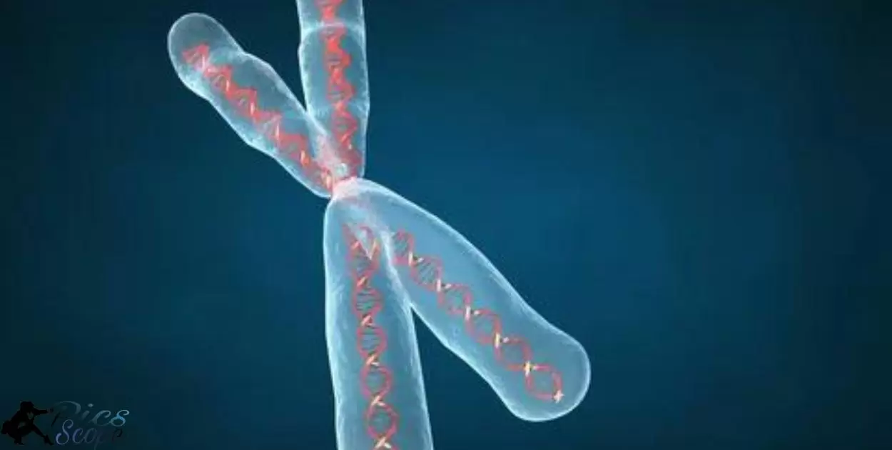
A photograph of an individual’s chromosomes shows the chromosome makeup and DNA of that person. Chromosomes are threadlike structures within cells that carry genetic information in the form of DNA. Humans have 23 pairs of chromosomes. A karyotype is the photograph produced when chromosomes from a cell are stained and arranged by size, number, and shape.
A Photograph Of An Individual’s Chromosomes” reveals intimate details about a person. These threadlike strands dictate everything from hair color to disease risk. A single photo can unlock generations of family ancestry and traits. A Photograph Of An Individual’s Chromosomes becomes a map to the very origins of a human life.
A karyotype offers a glimpse into the fundamental building blocks of life. Chromosomes and the DNA they contain provide the instructions for how the body develops, inside and out. Examining A Photograph Of An Individual’s Chromosomes” allows scientists to identify abnormalities and study how DNA works.
Is Chromosome Photography Essential For Understanding Genetics?
Chromosome photography, also called karyotyping, allows scientists to visualize and analyze an organism’s chromosomes. This provides a genome-wide snapshot that reveals chromosomal alterations indicative of genetic diseases. Karyotyping combines microscopy and photography to capture chromosome images that are arranged into informative karyograms.
Banding techniques stain chromosomes to produce unique banding patterns. These distinctive bands help identify individual chromosomes and diagnose defects like breakage, loss, duplication, translocation or inversion. Karyotyping is therefore an essential cytogenetic technique for understanding genetics.
How Does Photographing Chromosomes Reveal Genetic Traits?
Karyotyping allows chromosomes to be paired and ordered to detect abnormalities. The banding patterns help correlate DNA sequence data from the Human Genome Project with visual cytogenetic bands. This facilitates identifying candidate genes within implicated chromosomal intervals.
Photographing chromosomes also aids studying chromosomal translocations. Spectral karyotyping assigns pseudo-colors to distinguish individual chromosomes. Observing translocations reveals genetic alterations tied to genetic traits and disorders.
What Diseases Can Be Detected From Chromosome Photography?
Several genetic diseases manifest physically as chromosomal abnormalities that karyotyping can detect. For example, Down syndrome stems from an extra copy of chromosome 21. Klinefelter syndrome involves an additional X chromosome in males. Chromosome 15 deletions cause Prader-Willi syndrome imprinting disorders.
Cancerous cells often exhibit chromosomal defects. The Philadelphia chromosome translocation mutation is commonly associated with chronic myelogenous leukemia. Karyotyping thus aids diagnosing numerous diseases.
Can Photographs Of Chromosomes Predict Lifespan?
Telomeres are repetitive sequences at chromosome ends that protect DNA integrity. Shortened telomeres can indicate cellular aging and are associated with reduced lifespan. Techniques like quantitative fluorescence in situ hybridization (Q-FISH) can directly visualize and measure telomere length from chromosome photographs.
However, telomere length is not an absolute predictor of lifespan. Long telomeres don’t guarantee longer lifespans, although short ones correlate to mortality risk. Karyotyping alone cannot definitively predict lifespan, but it provides clues about biological aging.
What Are The Limitations Of Chromosome Photography?
Chromosome photography has resolution limits. It cannot detect small changes below 5 megabases. The images also depend on dye binding, which can introduce artifacts. Sample preparation is challenging and can alter structure.
Electron microscopy offers higher resolution views of chromosome structure. But it still cannot capture native state well due to condensation changes during metaphase. The sample preparation also affects structure. Transmission methods have additional limitations.
Does Chromosome Photography Violate Privacy?
Chromosome images contain genetic information. So privacy concerns can arise from accessing or sharing these photos without consent. Research studies likely obtain proper permissions and ethics approvals for using chromosome images.Clinical testing also requires patient consent. So violations seem unlikely in legitimate contexts. Karyotype analysis examines chromosomes to detect abnormalities associated with disorders. The Photograph Help Support The Report’s Descriptions
Can Photographs Of Chromosomes Be Inaccurate?
Yes, chromosome images can be inaccurate. The binding of fluorescent dyes introduces artifacts that alter structure. And the condensation state during metaphase changes native morphology.
Subjective analysis by technicians can also lead to misinterpretation. Automated analysis has improved accuracy but still has limitations. So false positives or negatives happen.
Is Photographing Chromosomes Expensive And Time Consuming
Chromosome imaging requires many steps like cell culture, staining, microscopy. So it is relatively expensive and slow. Sample preparation takes weeks in some cases. Analysis time also lengthy as technicians must visually inspect images. Automation helps but still needs review. So process from start to finish consumes significant time and resources.
How Has The Technology For Photographing Chromosomes Evolved?
Early chromosome photography relied on basic light microscopes and manual photography techniques. Researchers had to stain the chromosomes and then quickly take a picture before the sample dried out or faded. Over time, fluorescent stains, digital cameras, and computer-controlled microscopes have automated and improved the process.
Modern chromosome imaging utilizes advanced fluorescent tags, high resolution digital cameras, and sophisticated software to capture, process, and analyze images. Digital systems provide more control, speed, and accuracy for photographing chromosomes. Researchers can now instantly view, save, and share chromosome images globally.
How Have Camera Improvements Enhanced Chromosome Photography?
Early chromosome photos were black and white and low resolution due to film technology limitations. Images lacked fine detail needed for analysis. Modern digital cameras offer vastly improved resolution and color imaging capabilities ideal for detecting subtle chromosome features.
Digital photography also enables instant viewing of images. Researchers can immediately review photos to check quality rather than waiting for film development. They can quickly adjust microscope settings on the fly to refine images rather than losing precious preparation time.
How Have Staining Techniques Advanced Chromosome Photography?
Original chromosome stains like Giemsa offered basic differentiation of chromosome bands. New fluorescent tags precisely highlight specific chromosome regions and components like genes or telomeres. These tags emit signals detectable with digital cameras but invisible to the human eye.
Multiple fluorescent tags labeling different targets can now be used together. This allows simultaneous visualization of several chromosome elements in full color images. Advanced stains better differentiate chromosome shape and structure to enhance analysis.
How Has Image Processing Software Impacted Chromosome Photography?
Chromosome analysis originally required manual inspection of photos. Modern software can digitally enhance, measure, and compare chromosome images to detect subtle abnormalities.
Image processing algorithms can sharpen chromosome detail, overlay multiple photos, and extract quantitative data on chromosome shape or banding patterns.
Automated karyotype assembly software can arrange and number chromosomes to streamline genetic mapping. Digital systems provide more objective and nuanced chromosome classification.
What New Chromosome Photography Techniques Are Emerging?
X-ray ptychography allows imaging chromosomes without lenses or dyes. This uses X-ray photons to interact with atoms in the sample. Another emerging method is 3D imaging that connects many loci along DNA chains to form a comprehensive picture.
Researchers are developing new computational techniques like computer vision to analyze hundreds of chromosome images automatically. This can accelerate genetic analysis.
Can 3D Renderings Improve Chromosome Photography Analysis?
Yes, 3D renderings provide more accurate and detailed representations of chromosome structure. Researchers have captured high-resolution 3D images showing how chromatin folds.
Analyzing 3D images can reveal relationships between karyotype structure and function. Researchers are building 3D maps of all 46 chromosomes, which will enable better understanding of mechanisms underlying organization.
Could AI Enhance Chromosome Photography Interpretation?
Yes, AI could help automate analysis of chromosome images to accelerate genetic testing. Techniques like computer vision are already being applied to extract more insights from photographs.
As more high-resolution chromosome images are collected, machine learning algorithms can be trained on them. This could aid diagnosis of abnormalities and personalized medicine.
Could Smartphone Cameras Democratize Chromosome Photography?
Perhaps in the future, smartphone cameras and computational techniques could enable rapid chromosome analysis outside laboratories. This could make genetic testing more accessible and widespread. Current smartphone cameras likely lack sufficient resolution for detailed chromosome imaging.
What Does The Future Hold For Photographing Chromosomes?
Advances in cryogenic technology will enable imaging chromosomes in their native state without harsh chemical fixation. This could reveal new insights into chromosome structure and function. Staining methods like DNA painting may also improve resolution. Overall the future is moving towards full cryo imaging for studying fundamental properties of chromosomes.
Will Genome Sequencing Replace Chromosome Photography?
Genome sequencing provides base pair level detail but lacks spatial context. Imaging maps chromosome structure and spatial relationships critical for gene regulation. Both methods remain important. Sequencing assembled reference genomes enables targeted imaging of genomic loci along chromatin.
Could Live Cell Chromosome Photography Be Possible?
Live cell imaging faces challenges like diffraction limits and photo-toxicity. But advances like expansion microscopy show promise for less invasive imaging with higher resolution. Machine learning can also enhance live imaging. So live cell chromosome movies seem feasible in the future.
Will Public Chromosome Photography Databases Emerge?
As imaging and sequencing data accumulates, public databases would enable data sharing and reuse. Standardized datasets with rich metadata would also allow training machine learning algorithms. Given the value demonstrated in genomics, similar chromosomal atlases could emerge as methods and data mature.
FAQ’s
What do you call a picture of someone’s chromosomes?
A karyotype.
What is the name for a visual display of a person’s chromosomes?
Karyogram.
What term refers to a photograph showing the chromosomes of an organism?
Karyotype.
What are images depicting an individual’s chromosomes known as?
Karyotypes.
What is the technical name for a photographic representation of the chromosomes from a single cell?
A karyotype is the term for such a photographic depiction of an individual’s chromosomes.
Conclusion
A karyotype is a photograph of an individual’s chromosomes. The chromosomes are arranged in pairs by size and shape. Scientists take pictures of chromosomes to study them.
Karyotypes help detect chromosome abnormalities.
These abnormalities cause genetic disorders. Scientists use karyotypes to diagnose disorders. Patients consent for doctors to take karyotype pictures. Privacy of genetic data is protected by laws. Karyotyping is an important genetic test.