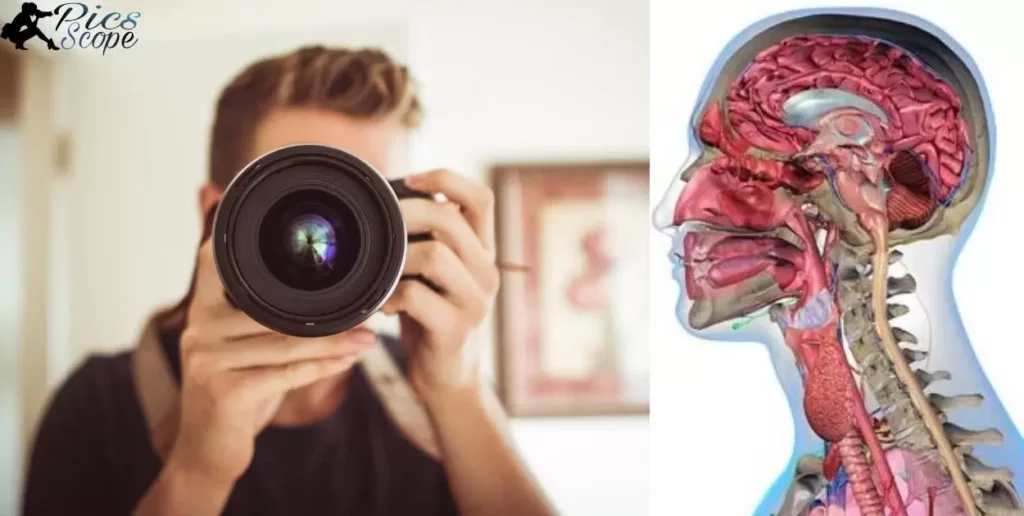
Photography is the art of capturing images with light. In A Photographic Atlas For Anatomy & Physiology, it visually documents the complexities of the human body, offering a clear understanding through the lens.
A Photographic Atlas For Anatomy & Physiology is not just a book; it’s a visual gateway to the intricacies of the human body. Using the power of photography, this atlas transforms learning into an engaging exploration that goes beyond traditional textbooks.
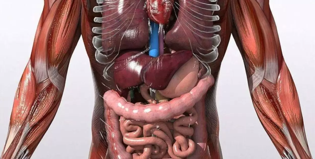
Within its pages, A Photographic Atlas For Anatomy & Physiology combines anatomical precision with photographic artistry. Each carefully curated image serves as a snapshot, revealing the mysteries of anatomical structures and physiological processes, making it an indispensable resource for students, educators, and enthusiasts.
What Role Does Photography Play in A Photographic Atlas For Anatomy & Physiology?
Photography in A Photographic Atlas For Anatomy & Physiology serves a crucial purpose by visually presenting intricate details of the human body. It acts as a tool to simplify complex concepts.
Offering a clear and tangible representation of anatomical structures and physiological processes. Through straightforward visual communication, it aids students and enthusiasts in better understanding the complexities of anatomy.
This approach transforms learning into an engaging exploration, where each photograph is a carefully chosen snapshot that vividly captures the essence of anatomical features.
In this way,A Photographic Atlas For Anatomy & Physiology utilizes the power of photography to make the study of anatomy accessible and intriguing, providing an indispensable resource for those eager to delve into the wonders of the human body.
Exploring the Artistic Merits of Photography in Anatomy Education
In anatomy education, Course In Photography Digital 4th Edition Pdf brings a unique artistic touch. It turns complex structures into visual stories, making learning engaging and accessible. Through vivid images, students discover the intricacies of the human body, fostering a deeper understanding.
The artistic merits of photography shine in A Photographic Atlas For Anatomy & Physiology. This resource transforms anatomy education by using visual elements. Students benefit from the clarity of images, turning the study of anatomy into a visually compelling experience.
How Does A Photographic Atlas For Anatomy & Physiology Enhance Visual Learning?
In A Photographic Atlas For Anatomy & Physiology, pictures play a crucial role in making learning more visual and engaging. This book uses photography to show detailed images of anatomical structures, helping students understand complex concepts easily.
The visual approach makes it easier for learners to grasp the intricate details of the human body. Instead of lengthy descriptions, the atlas relies on clear, concise images, allowing students to absorb information quickly and retain a better understanding of anatomy and physiology.
Unveiling the Intricacies, Photography’s Impact on Anatomy Understanding
In A Photographic Atlas For Anatomy & Physiology, photography plays a crucial role in helping us understand the complexities of the human body. It captures detailed images, making it easier for students and enthusiasts to grasp anatomical structures.
These visuals act as a bridge, simplifying the learning process by providing a direct and clear representation of intricate physiological details. Through photography, the impact on anatomy understanding is immediate and tangible.
The visual nature of this approach eliminates confusion and enhances comprehension, allowing individuals to explore and appreciate the nuances of the human body. In essence, the use of photography in this context transforms the study of anatomy into an engaging and accessible experience for learners at all levels.
Can Photography Truly Capture the Essence of Human Physiology?
In A Photographic Atlas For Anatomy & Physiology, photography becomes a tool for revealing the intricacies of the human body. It visually captures the essence of human physiology, presenting a clear and direct portrayal through carefully chosen images.
The visual impact of photography in this atlas ensures that the intricacies of anatomical structures are accessible to all learners. It’s a straightforward approach that allows readers to engage directly with the subject matter, making the study of human physiology more tangible and relatable.
The Intersection of Science and Art, Photography in Anatomy Studies
The intersection of science and art in Photography in Anatomy Studies melds precision with visual storytelling, offering a unique lens into the intricacies of the human body. Through carefully captured images, this fusion harmonizes scientific exploration with aesthetic elements.
Simplifying complex anatomical concepts for an engaging and accessible learning experience. In this dynamic blend, photography transcends traditional boundaries, transforming anatomical communication into a visually compelling narrative
Harmony of Form and Function,
Photography in anatomy studies creates a unique intersection between science and art, harmonizing the precision of scientific exploration with the aesthetic elements of visual storytelling.
Each image in A Photographic Atlas For Anatomy & Physiology carefully captures the form and function of anatomical structures, transforming scientific data into visually compelling narratives.
Visualizing Complexity with Simplicity,
The use of photography simplifies the complex subject matter of human anatomy, presenting it in a way that is easily digestible for learners. Through visually striking images, intricate physiological processes become more approachable, breaking down barriers to understanding and allowing for a seamless integration of science and art.
Aesthetic Accuracy in Anatomical Communication,
Photography in anatomy studies serves as a tool for accurate and aesthetically pleasing communication of anatomical knowledge. The carefully curated images not only convey scientific information but do so in a visually engaging manner, making the learning experience more enjoyable and effective for students and enthusiasts alike.
Capturing Dynamics in Motion,
Unlike traditional static illustrations, photographic images can capture the dynamics of physiological processes in real-time. This dynamic quality adds a layer of depth to the study of anatomy, allowing learners to visualize movement and function, fostering a more comprehensive understanding of the human body.
Democratizing Access to Knowledge,
The intersection of science and art through photography in anatomy studies plays a crucial role in democratizing access to anatomical knowledge. Visual representation transcends language barriers.
Making the study of anatomy more inclusive and accessible to a diverse audience, irrespective of their scientific background. This harmonious blend of science and art in the form of photography ensures that the beauty and complexity of the human body are appreciated and understood by all.
How Does the Use of Photography Elevate Anatomical Education?
Photography in anatomical education makes learning clearer and more engaging. Instead of just reading about structures and processes, students see detailed images that bring concepts to life. These visuals enhance understanding by providing a direct and visual connection to the intricacies of the human body.
In A Photographic Atlas For Anatomy & Physiology, photography takes center stage as a tool for effective teaching. Students benefit from a more direct and concrete experience, allowing them to grasp anatomical details in a way that textbooks alone cannot achieve.
Photography in Anatomical Communication
- Visual Clarity, Photography in anatomical communication enhances clarity by providing clear, detailed images of anatomical structures.
- Effective Teaching Tool, It serves as a powerful teaching tool, allowing educators to convey complex anatomical information more effectively.
- Enhanced Understanding, Students benefit from a visual language that goes beyond traditional methods, facilitating a deeper and more intuitive understanding of anatomy.
- Communication Bridge, Photography acts as a bridge in anatomical communication, breaking down barriers and making intricate concepts more accessible.
- Detailed Representation, The visual language of photography captures the nuances of anatomical features, offering a comprehensive and detailed representation.
- Active Engagement, The use of photography encourages active engagement, making the learning experience more dynamic and memorable.
- Accessible Learning, It makes anatomical communication more accessible to a wider audience, catering to different learning styles and preferences.
The Significance of Visual Storytelling, Photography in Physiology
Photography in physiology is essential for clear communication. It simplifies complex concepts, allowing students to grasp details easily. Visual storytelling, as seen in A Photographic Atlas For Anatomy & Physiology, transforms the learning experience.
In this approach, images speak louder than words. Instead of relying on lengthy explanations, visual storytelling captures the essence of physiological processes in a glance. The significance lies in its ability to break down barriers, making physiology accessible to all through the power of vivid and engaging photographs.
How A Photographic Atlas For Anatomy & Physiology Transforms Learning Through Images
In A Photographic Atlas For Anatomy & Physiology, pictures become teachers. These images show body parts, making learning easier. Instead of just reading, you see and understand.
The atlas is like a visual guidebook. It uses photos to explain complex things. When you study with it, learning about the human body becomes a visual adventure.
Exploring the Educational Value of Photographic Representation in Anatomy
In Exploring the Educational Value of Photographic Representation in Anatomy, pictures play a crucial role in helping students learn about the human body. These images serve as a visual guide, making complex anatomical structures more understandable.
By incorporating photography, the educational experience becomes more engaging and accessible. Photographs in anatomy education offer a direct and straightforward way to grasp intricate details.
Instead of relying solely on written descriptions, students can see and identify anatomical features, fostering a more hands-on and visual approach to learning. With the use of photographic representation, the study of anatomy becomes not only educational but also visually stimulating and memorable.
What Sets Apart A Photographic Atlas For Anatomy & Physiology in Visual Education?
A Photographic Atlas For Anatomy & Physiology stands out by using clear images to teach about the human body. Unlike traditional textbooks, this atlas relies on the power of photography for an engaging learning experience.
In this book, each photo is carefully chosen to explain anatomical structures and physiological processes. The visual approach makes it easy for students to grasp complex concepts, turning the study of anatomy into a captivating journey through the lens of photography.
Unlocking the Wonders of the Human Body, A Photographic Journey

In A Photographic Atlas For Anatomy & Physiology, we embark on a visual journey that uncovers the wonders of the human body. The atlas uses photography to bring clarity and simplicity to the intricate structures and processes within our anatomy.
Through crisp and detailed images, readers gain a direct and tangible understanding of the complexities of the human body. This visual approach enhances learning, making it an engaging experience for students and anyone curious about the marvels of anatomy and physiology.
Photography’s Contribution to Clarity in Understanding Anatomical Structures
Photography in A Photographic Atlas For Anatomy & Physiology brings unparalleled clarity to understanding anatomical structures. Through vivid images, it offers a precise visual guide, simplifying complex concepts for enhanced comprehension. The contribution of photography ensures a clear and accessible exploration of the intricacies of the human body.
Visual Precision in Anatomy Education
Photography plays a pivotal role in enhancing the clarity of anatomical structures by providing a visual precision that textbooks alone cannot achieve. Through high-resolution images, A Photographic Atlas For Anatomy & Physiology allows learners to observe intricate details with clarity, facilitating a deeper understanding of the human body’s complex composition.
Accessible Learning through Visual Representation
The utilization of photography makes anatomical structures more accessible to a broader audience. With clear visual representations, this atlas breaks down complex concepts into easily digestible components, catering to various learning styles.
The visual precision offered by photography ensures that students, educators, and enthusiasts alike can grasp the intricacies of anatomical structures with ease.
Eliminating Ambiguity through Photographic Detail
Photography eliminates ambiguity in understanding anatomical structures by capturing details with precision. Each image in the atlas serves as a roadmap, guiding learners through the various layers and components of the human body.
Bridging the Gap Between Theory and Practice
In the realm of anatomy education, photography serves as a bridge between theoretical knowledge and practical application. By providing clear visual representations of anatomical structures, learners can connect textbook concepts to the real-world, tangible reality of the human body.
Enhancing Retention Through Visual Engagement
Photography’s contribution to clarity extends beyond understanding; it actively engages learners, thereby enhancing retention. The visual appeal of the images in the atlas creates a memorable learning experience.
Helping individuals retain and recall information more effectively. By combining clarity with visual engagement, photography becomes a powerful tool for fostering a lasting understanding of anatomical structures.
Can Photography Bridge Gaps in Anatomy Education for Students?
Photography plays a crucial role in helping students understand anatomy better. Instead of relying solely on words, images in A Photographic Atlas For Anatomy & Physiology act as visual guides, making complex structures more accessible. Students benefit from this hands-on approach, as it simplifies learning and enhances comprehension.
By incorporating photography, this atlas bridges gaps in anatomy education. Students can see detailed images of anatomical structures, aiding in memorization and understanding. The visual nature of photography makes the subject more engaging, transforming it from a theoretical concept into a tangible, visual experience for students.
How Photography Reshapes Traditional Anatomy Learning
Photography changes how we learn about anatomy. In traditional learning, words dominate, but with A Photographic Atlas For Anatomy & Physiology, images take the lead. This shift makes complex anatomical structures more accessible, allowing learners to grasp concepts quickly.
Visual impact is crucial in anatomy education. A Photographic Atlas For Anatomy & Physiology uses photography to bring clarity and precision to the study of the human body. Instead of relying solely on text, the visual emphasis reshapes the learning experience, making it more engaging and effective for students.
What Visual Insights Does A Photographic Atlas For Anatomy & Physiology Offer?
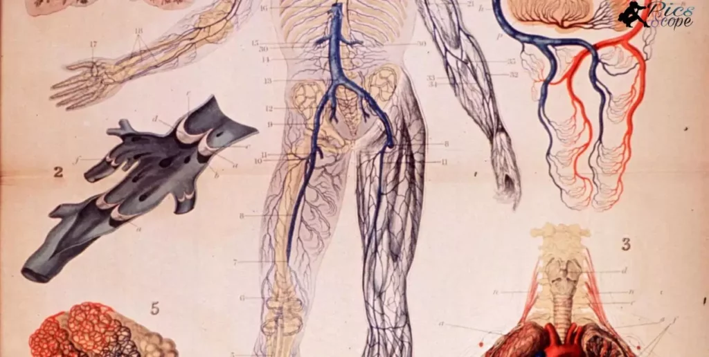
- Detailed Visuals, The atlas provides intricate images that capture the nuances of anatomical structures.
- Clarity in Complexity, Visual insights help simplify complex physiological processes for easy understanding.
- Comparative Anatomy, Side-by-side comparisons enhance comprehension of variations in anatomical features.
- Real-world Context, Photographs offer a realistic portrayal, bridging the gap between theory and practical application.
- Sequential Presentation, Visual sequences aid in understanding processes and the interconnectedness of body systems.
- Pathological Illustrations, The atlas includes visuals of various pathologies, aiding in the recognition of abnormal conditions.
- Cross-Sectional Views, Different perspectives and cross-sections offer a comprehensive understanding of structures.
- Clinical Relevance, Visual insights highlight practical applications in clinical settings, connecting theory to practice.
- Diversity of Cases, The atlas covers a broad spectrum of cases, providing a well-rounded visual learning experience.
- Enhanced Memorization, Visual stimuli contribute to better retention and recall of anatomical details.
- Anatomical Variations, Images showcase variations in anatomy, preparing learners for real-world diversity.
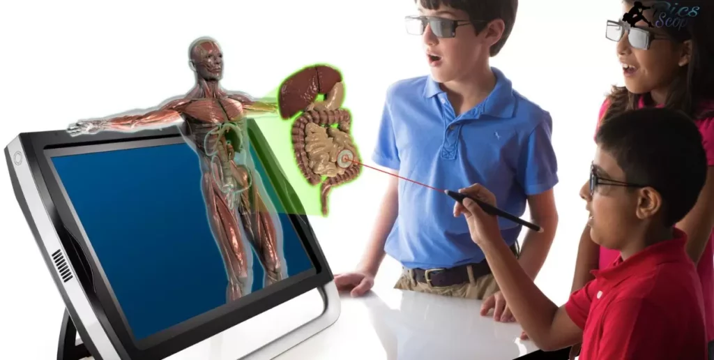
- Interactive Learning, Visual insights encourage active engagement, fostering a deeper understanding of anatomy.
- 3D Visualizations, Some images offer a three-dimensional perspective, enhancing spatial understanding.
- Microscopic Details, Close-up visuals reveal microscopic details, aiding in the study of cellular anatomy.
- Anatomical Relationships, Visual insights emphasize the relationships between different anatomical structures.
- Systematic Organization, The atlas organizes visuals in a systematic manner, facilitating step-by-step learning.
- Surgical Perspectives, Some visuals provide insights from a surgical standpoint, linking theory to medical practice.
- Developmental Stages, Visuals depict anatomical structures at various developmental stages, offering a comprehensive view.
- Physiological Processes, Images illustrate dynamic physiological processes, enhancing understanding beyond static diagrams.
- User-Friendly Format, The visual presentation is designed for easy navigation and quick reference.
How Photography Enhances the Accessibility of Anatomy & Physiology Education
Photography makes learning anatomy and physiology more accessible by providing clear visual representations. In A Photographic Atlas For Anatomy & Physiology, images serve as a practical tool, breaking down complex concepts into easy-to-understand snapshots.
Students benefit from the straightforward visuals, aiding their comprehension of intricate anatomical structures. The use of photography in anatomy education eliminates potential confusion that may arise from lengthy descriptions. Instead of relying solely on words, students can rely on the clarity of visual aids, making it easier to grasp and remember essential concepts.
Photography’s Precision in Anatomical Illustration
In A Photographic Atlas For Anatomy & Physiology, photography is like a close-up lens, showing precise details of the human body. The pictures capture the intricacies of anatomical structures in a way that’s easy to understand. Each image is a clear, straightforward illustration, making learning anatomy more accessible.
The precision of photography in anatomical illustration is striking. It brings the details to the forefront, making it easier for students to grasp complex concepts. With each close-up shot, the atlas becomes a visual guide, breaking down the barriers to understanding anatomy and physiology.
Key Element in A Photographic Atlas For Anatomy & Physiology
Photography is crucial in A Photographic Atlas For Anatomy & Physiology. It’s not just about pictures; it’s a practical tool enhancing understanding. The images, carefully chosen, serve as snapshots, making anatomy and physiology accessible.
In this atlas, photography plays a vital role. It’s not just an art; it’s a practical and key element. Through clear visuals, readers gain a practical grasp of the intricate details of the human body.
FAQ’s
What does A Photographic Atlas For Anatomy & Physiology” offer?
This atlas provides a visual journey through the intricacies of the human body, using photography to enhance understanding.
How does photography contribute to anatomy education in this atlas?
Photography in the atlas serves as a practical tool, offering clear visuals that make anatomical details accessible and engaging.
Why is the practicality of photography emphasized in this context?
Emphasizing the practicality of photography underscores its role in enhancing comprehension, providing a tangible and visual approach to learning anatomy and physiology.
Can this atlas be useful for students and educators alike?
Absolutely, “A Photographic Atlas For Anatomy & Physiology” serves as an invaluable resource for students, educators, and anyone interested in exploring the marvels of the human body.
What sets apart this atlas from traditional anatomy textbooks?
This atlas stands out by leveraging the power of photography, offering a unique and captivating perspective that goes beyond traditional textbooks, making the learning experience more dynamic and engaging.
Conclusion
In conclusion, ,,A Photographic Atlas For Anatomy & Physiology,, proves to be an invaluable resource for learners. The integration of photography brings a practical dimension to studying the complexities of the human body, making it a key element in the educational journey.
By merging the precision of anatomical science with the artistry of photography, this atlas offers a unique and accessible way to understand anatomy and physiology. In the realm of visual education A Photographic Atlas For Anatomy & Physiology stands out as an indispensable tool, providing a clear and practical lens through which learners can explore the wonders of the human body.