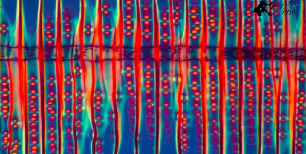
A photographic atlas is a collection of photographs and images that systematically illustrate a specific subject. In the context of developmental biology, a photographic atlas would contain images depicting the various stages of development of organisms. The images aim to provide a visual guide to understanding developmental processes.
A Photographic Atlas of Developmental Biology” immediately conjures vivid images of embryos, fetuses, and organisms at different phases of growth. Such an atlas promises to be a visual feast, allowing readers to witness firsthand the miraculous progression from a single cell to a fully formed creature.
An atlas focused specifically on developmental biology would serve as an invaluable educational resource. By compiling photographs and illustrations, it would enable students and researchers to directly compare the developmental milestones across various organisms. Such comparative analyses can reveal common patterns and mechanisms underlying embryogenesis, organ formation, and growth to maturity.
What Does Photographic Developmental Biology Photography Capture?
Photographic developmental biology aims to visually document the progression of organisms from conception through maturation. Photographs capture intricate developmental phenomena that words struggle to describe, from the earliest stages of embryogenesis through complex morphogenetic movements involved in organ formation.
Images freeze fleeting milestones, allowing detailed study. Photography also facilitates comparative analyses across organisms. Similarities in developmental programs become apparent when conserved embryonic features are captured side-by-side. Subtle differences also emerge upon close visual inspection. Comparative photographic atlases can reveal both unity and diversity in the developmental biology of life.
How Does Embryo Photography Illustrate Developmental Stages?
Embryo photography provides vivid illustrations of early developmental events. Sequential images can trace cell cleavage patterns starting from the fertilized zygote through blastulation and gastrulation. Photomicrography reveals cellular migrations involved in germ layer formation and early body patterning.
Photographing live embryos poses technical challenges but offers scientific payoffs. Advances in non-invasive imaging techniques now enable tracking cell lineages, monitoring gene expression, and visualizing physiological processes without disturbing normal development. As imaging tools improve, embryo photography will continue generating insights into the intricate dance of developmental biology.
What Cellular Processes Are Highlighted in Microphotography?
Microphotography utilizes microscopes to capture developmental biology at the cellular and subcellular scales. Photomicrographs visually accentuate intricate mechanisms involved in cell proliferation, migration, and differentiation.
Time-lapse imaging can condense these dynamic processes, showing mitotic cell divisions, cytoskeletal rearrangements guiding motility, and morphogenetic cell shape changes underlying tissue morphogenesis.
Fluorescence macro photography spotlights specific molecules and structures tagged with light-emitting labels, quantitatively mapping their changing distribution through space and time. Photomicrographs also form the basis for computational analyses of cell behaviors.
Can Time-Lapse Photography Reveal Growth Patterns?
Time-lapse photography compresses extended developmental sequences into compact visual animations, facilitating analysis of growth dynamics. Condensing hours or days of embryogenesis into minutes of time-lapse footage accentuates patterns of morphogenesis. Repeated imaging captures transient structures and illuminates complex cell behaviors underlying tissue movements in real time.
Modern time-lapse imaging also enables long-term monitoring of growth, documenting post-embryonic developmental changes throughout juvenile stages or even an organism’s entire lifecycle. This growth data can be correlated with genetic and environmental variables to uncover factors regulating developmental timing, size, and form.
How Can Photomicrography Enhance Understanding of Embryogenesis?

Photomicrography, the practice of photography through a microscope, can greatly enhance scientific understanding of embryogenesis. High magnification imaging allows developmental biologists to visually track developmental progress on a cellular and even molecular scale.
Photomicrographs can capture intricate details of cell division, migration, and differentiation as embryos transition through key growth phases. Comparison of images across embryo samples reveals both consistencies as well as variations in early development.
Advanced photomicrography techniques, such as fluorescence and confocal microscopy, enable even clearer visualization of embryonic development. Fluorescent labeling of specific cells or structures lets researchers follow their precise movements and interactions over time. Confocal microscopy facilitates optical sectioning through thick embryo samples, allowing construction of 3D models of embryo architecture during morphogenesis.
What Are Key Embryo Development Milestones Captured Through Photography?
Photography has been instrumental in documenting major milestones in embryo development across species. Images can definitively pinpoint stages such as gastrulation, neurulation, and organogenesis by capturing morphological landmarks.
Photography also enables measurement of growth variables like embryo length, somite number, and limb bud dimensions to quantitatively assess developmental progress. Time-lapse photographic sequences beautifully demonstrate dynamic processes like neural tube closure and heart looping.
At the cellular level, photomicrographs reveal milestones like egg activation after fertilization, blastocyst formation, and axis specification during gastrulation. Photography has also been essential for characterizing abnormal development in embryos with genetic or environmental defects.
Overall, photographic atlases systematically link embryo morphology at precise timepoints to known genetic and molecular markers of key developmental transitions. Thus images provide visual benchmarks that researchers utilize to evaluate embryo development in diverse studies.
How Does Photographic Mapping of Cell Lineages Aid Research?
The ability to track cell divisions and migrations is crucial for elucidating mechanisms controlling embryo growth. Photographic mapping of cell lineages involves labeling individual cells, usually with fluorescent markers, and then microphotographing embryo samples over time.
This generates visual cell fate maps depicting the sequence and timing of cell divisions, movements, and differentiation events as development proceeds. Researchers can also photographically compare cell lineage patterns between normal and genetically perturbed embryos to clarify roles of specific genes.
Photographic mapping data complements genetic fate mapping techniques for assembling a comprehensive understanding of embryogenesis. Cell lineage analyses are also essential for studies focused on tissue regeneration and stem cell based organoids.
What Insights Does Comparative Embryo Photography Provide?
Comparative embryo photography, imaging embryos across diverse species, can identify common developmental blueprints across phylogeny. Similar sequences of morphological changes during embryogenesis argue for conserved genetic and morphogenetic mechanisms. Photography Was Invented
Variations can also be highly informative. For example, photographic comparisons of marsupial and eutherian mammal embryos revealed that marsupial neonates are born at an earlier developmental stage more equivalent to eutherian embryos. This suggests fundamental differences in their reproductive strategies.
Photographic atlases that systematically document and compare embryonic development across taxa have proven invaluable resources for evolutionary developmental biology. Images enable quick visual appraisal of similarities and differences in growth patterns, facilitating quantitative analyses and experimental comparisons between species.
What Standards Guide Assembly of Developmental Biology Image Collections?
Photographic atlases aimed at research and educational applications adhere to strict standards for image quality and documentation. Factors like microscope magnification, staining methods, and embryo staging must be reported. Images are also evaluated for resolution, color accuracy, contrast, and field of view. Standardized protocols allow appropriate comparison across species.
For example, the Zebrafish Atlas Project assembles images using agreed-upon developmental staging. Compliance with data standards facilitates integration with genetic and genomic repositories.
Image collections are further subject to ethical considerations regarding specimen acquisition and photographic techniques. Journals and atlases must confirm appropriate animal use oversight and permits were obtained. Digital techniques like computational stitching demand additional scrutiny regarding manipulation.
How Are Photographic Atlases Organized for Educational Use?
Photographic atlases are designed to align with pedagogical goals in developmental biology curricula. Introductory atlases categorize images by developmental stage, allowing students to directly compare embryonic and fetal morphology across organisms. More advanced compilations organize by tissue type and organ system, elucidating cellular dynamics underlying specialization and maturation.
Interactive digital atlases further sort images by researcher annotations and user tags. Such organization enables instructors to craft targeted image sets for lectures and labs focused on key developmental events like neurulation or limb formation. By systematically indexing photographic content to core learning objectives, atlases transform static images into versatile teaching tools that promote lasting comprehension.
What Digital Platforms Increase Accessibility of Image Atlases?
Online open access repositories have expanded availability of specialized image collections well beyond the shelves of university libraries. Leading developmental biology journals have created dedicated portals archiving high-resolution photo atlases indexed to published research.
Institutional image libraries further aggregate content, often enhanced by 3D visualization and multi-layer annotation tools. Most significantly, the proliferation of internet search engines now allows any student to instantly locate a relevant embryonic image based on species and developmental age.
While digital dissemination vastly increases access, ethical obligations persist regarding appropriate image use and attribution. Responsible citation and copyright adherence must feature prominently in any online platform to uphold scientific integrity.
What Are Limitations of Visual Representations in Developmental Biology?
Visual representations like diagrams and photographs are widely used in developmental biology to depict complex dynamic processes. Static images have limitations in conveying the full complexity of developmental phenomena that unfold over both space and time.
Simplified representations may reduce motivation to fully explore a concept if the visuals are not aesthetically engaging or optimally informative. There are also risks of promoting misconceptions if key details are left out.
Can Static Images Convey Dynamic Development Processes?
A key challenge is that static images struggle to capture the dynamic transformations that occur during development. Processes like cellular differentiation and morphogenesis involve complex spatiotemporal coordination across tissues and structures.
Still images lack dimensionality and can flatten critical aspects of developmental phenomena. Animations and interactive models may be better suited, but come with greater production costs.
Do Simplified Images Risk Promoting Misconceptions?
In an effort to clarify complex concepts, textbook illustrations often utilize simplifying abstractions by omitting real-world details. Excessive simplification runs the risk of promoting student misconceptions about developmental processes. Without sufficient contextual cues, students may over-generalize or form incorrect mental models.
How Can Captions Enhance Interpretation of Developmental Photography
When using actual photographs of embryo development, captions play a vital role in guiding student interpretation. For example, indicating specific anatomical structures, developmental stages, and orientation within the overall organism can help students form an accurate mental framework. Thoughtful captions are key for transforming static images into more meaningful visual explanations
How Can Photography Complement Quantitative Analyses in Studying Growth?

Photography provides visual documentation and qualitative data to complement quantitative measurements over time. Time-lapse or stop-motion photography can capture dynamic processes like growth that are difficult to quantify at fine time scales.
Images show spatial patterns and morphological changes that numbers alone may not convey. Comparisons of images over time also enable quantitative analysis of growth rates, proportions, etc. Overall, photography contextualizes, validates, and enhances quantitative data analysis.
How Can Images Calibrate Computational Models of Development?
Images of morphology at progressive developmental stages can be used to set parameters and test predictions of computational models that simulate growth. The visual benchmarks that images provide help constrain and validate simulations to match real empirical observations.
Images also suggest new quantitative metrics to characterize complex morphology that models attempt to recapitulate. Iteratively fitting models to morphological images enables refined modeling of developmental mechanisms.
What Are Best Practices for Integrating Photography with Genomic Data?
Linking images to genomic and molecular data facilitates identifying genetic regulators of visible phenotypes. Controlled imaging of mutants, knockdowns, etc. paired with omics profiling of the same samples enables connecting genetic and molecular signatures to morphological outcomes.
Images should capture informative views at consistent orientations and scales to quantitatively correlate morphology with genomic variables. Data management systems that associate images with multi-omics data sets empower integrated analysis.
How Can Images Enhance Mathematical Descriptions of Morphogenesis?
Photographic time series of developmental processes allow extracting quantitative measurements to inform parameters for mathematical models of morphogenesis. Images also provide qualitative validation of mathematical predictions for spatial patterning and morphological change over time.
Computational analysis of images, like segmentation and tracking, can derive spatial patterns to motivate new theoretical descriptions. Overall photographic documentation and quantification strengthens mathematical formalization of the physical mechanisms governing form and growth.
FAQ’s
What is a photographic atlas of developmental biology?
A photographic atlas of developmental biology contains images showing the progression of embryonic development across various species to educate students on morphological changes over time.
What types of photos are included?
Photos include microscopic and macroscopic images of embryos at progressive stages to illustrate anatomical structures, cell movements, and growth patterns during early development.
How can the atlas complement a textbook?
The visuals provide qualitative documentation of dynamic developmental processes described quantitatively in textbooks, strengthening comprehension.
What courses utilize such an atlas?
Typical courses are developmental biology, embryology, anatomy, and cell biology where understanding early morphological development is fundamental.
What analysis can students practice with the images?
Students can observe and compare embryonic stages across organisms, annotate structural changes, quantify growth rates, and relate form to genetic factors.
Conclusion
A Photographic Atlas of Developmental Biology serves an important role in education by providing visual documentation of key developmental processes. The photographic images act as valuable supplements to standard developmental biology textbooks.
They give students direct visualizations of complex morphological changes that are difficult to convey in words or diagrams alone. Having photographic benchmarks enriches the learning experience and comprehension of developmental concepts.
Photographic atlases will continue to be useful pedagogical tools for engaging students in developmental biology coursework. The visual and qualitative nature of photographic representations complements quantitative analysis and modeling approaches also used to study embryonic development.


